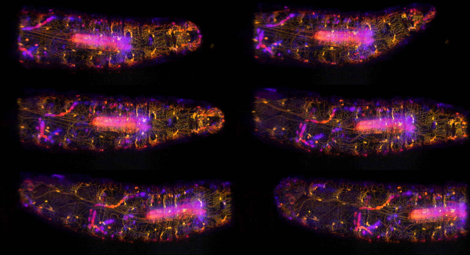
Image credit | Hillman Lab
Optical Biology 2023 Seminar Series
The online Optical Biology Seminar Series is run by our students. We invite scientists at the forefront of their field, who are developing and applying novel optical techniques to tackle biological questions. The talks are aimed at postgraduate students and postdoctoral researchers, both within and outside of UCL. For access to the talk recordings (when available), or any other inquiries, please email us at opticalbiology.seminars@gmail.com. We hope to see you there!
Wednesdays 5-6pm BST on Zoom
Register here
29 March |Molecular tools for studying the brain
Adam Ezra Cohen | Harvard University
Abstract Neurons communicate via electrical impulses, but until recently these signals have been invisible. Engineered opsin proteins from diverse microorganisms can, when expressed in neurons, convert the electrical activity of the cells into flashes of fluorescence. I will describe molecular, optical, and computational tools for imaging electrical signals in cultured neurons, brain slices, and behaving mice, and discuss applications to decoding neural circuit function. I will then describe some new molecular "tickertape" tools which open the possibility of whole-brain neural recordings.
12 April |TBC
Carolina Mora Lopez | Interuniversity Microelectronics Centre
7 June |Injury induces wild-type cell proliferation to suppress oncogenic Ras cells captured by intravital two-photon imaging
Sara Gallini | Yale University
Abstract Healthy skin is a tapestry of wild-type and mutant clones. Although injury can cooperate with Ras mutations to promote tumorigenesis, the consequences in genetically mosaic skin are unknown. Progress had been stymied by the inability to follow cell competition dynamics during skin repair after injury. To address this challenge, I pioneered an imaging approach that leverages the two-photon intravital microscopy. This approach allows me to track the same cells over time in live mice, and to investigate the consequences of injury-repair on genetically mosaic skin. I discovered that the presence of wild-type cells in Ras-mosaic skin suppresses the aberrant growth induced after injury. Moreover, whereas Ras-mutant cells outcompete wild-type cells in uninjured mosaic tissue, after injury their expansion is prevented due to a selective increase in wild-type cell proliferation. Epidermal Growth Factor Receptor (EGFR) ligands are known to activate Ras signaling, and I found that differential activation of the EGFR pathway and of proliferation in wild-type but not in Ras-mutant cells after injury explains the competitive switch. Pharmacological or genetic inhibition of EGFR abolished the proliferative advantage of wild-type cells after injury, restoring the expansion of Ras-mutant cells. Overall, I found that injury-repair unexpectedly switches the competitive advantage from Ras-oncogenic cells to wild-type cells in mosaic skin. This discovery reveals an unanticipated role for proliferative wild-type cells as a first line of defense against tumorigenic stimuli such as injury in genetically mosaic, healthy skin epithelium.
14 June |Speeding in Microscopy & Image Analysis (Expanding Single-Cell and In-Vivo Imaging Applications)
Kevin Tsia | Hong Kong University
Abstract This talk will describe how the powerful integration of ultrafast imaging advanced microfluidics and accelerated image analytics have been unlocking new possibilities in large-scale imaging cytometry and high-speed in-vivo imaging. We will dive into a number of successful examples in rare cancer cell detection, immune-cell sub-typing, targeted-drug sensitivity prediction, and kHz multiphoton imaging of neural activity in awake mice. I will also discuss the ongoing challenges and the importance of fostering a collaborative "optical biology" community bringing together microscopists bioinformaticians and biologists/clinicians to ensure widespread adoption and accessibility of advanced (high-speed) microscopy techniques.
21 June |Imaging self-organization of MAPK signaling dynamics in the epithelium
Oliver Pertz | Institute of Cell Biology, University of Bern
Abstract Cells dynamically sense and respond to ever changing external stimuli through sophisticated signaling networks. Accordingly, signaling dynamics rather than steady states control fate decisions. For many signaling pathways, heterogeneous dynamic signaling states occur within distinct cells, explaining fate variability observed within a cell population. Measuring single cell signaling dynamics is therefore key to understand how cellular responses correlate with specific cell fate decisions. Here, we combine biosensor imaging, optogenetics and mathematical modelling to map how different MAPK signalling network circuitries fine tune ERK activity dynamics at the single cell level.
We apply these technologies to study fate determination system in collectives of MCF10A breast epithelial cells. In a process called epithelial homeostasis, this cell collective constantly senses the state of the epithelium, and reacts by spatially tuning survival and proliferation fates to ensure a critical cell density necessary for proper barrier function. Here, we observe two single-cell ERK signalling modes that consist either of stochastic pulses (in presence of growth factor stimulation), or of co-ordinated ERK waves across multiple cell layers that originate around apoptotic extruding cells (in absence of GFs or in presence of cytotoxic stress). We show that such ERK activity pulses provide a local survival signal for about 4 hours to the cells surrounding the apoptotic extruding cells, allowing to fine tune epithelial homeostasis at the cell population level. We show that this spatial mechanism of ERK activity waves ensures that a critical number of cells is maintained in the epithelium, even in the presence of strong external insults such a chemotherapy.
A higher complexity in spatio-temporal signalling is observed when these cells are grown as 3D epithelial acini that display a specific size and exhibit a lumen. This sequentially involves initial cell proliferation, followed by quiescence and triggering of apoptosis specifically in the inner cell mass to allow for lumen formation. We show that spatio-temporal self-organization of ERK frequency within this cell ecosystem serves as a mechanism to locally control proliferation, survival and apoptosis fates during acinus growth and lumen formation. Our results provide new insight into the self-organization of signaling dynamics in morphogenesis of complex multicellular structures.
5 July |A Hybrid All-Optical Method for Studying Grid Cell Circuit Connectivity
Weijian Zong | Norwegian University of Science and Technology
Abstract The medial entorhinal cortex (MEC) is believed to create an internal map of an animal's position, which helps with spatial navigation. This map consists of several types of spatially-tuned cells that are active in specific locations or movement directions, such as grid cells, border cells, object-vector cells, and head-direction cells. Grid cells, in particular, are a striking cell type that encode spatial information using a hexagonal and periodic firing pattern that is not influenced by external sensory input but instead must be generated by local circuit computation. Understanding how grid cells are generated is crucial to understanding the algorithms used for spatial encoding in the mammalian brain. The most powerful theoretical framework for explaining grid-firing generation is based on the idea of continuous attractor networks (CAN), where specific recurrent synaptic connectivity constrains the joint activity of grid cells to a low-dimensional manifold. However, the key prediction of CAN models, which states that there is stronger excitatory connectivity between functionally similar cells and weaker connectivity between dissimilar cells, has not been tested due to a lack of technology that can simultaneously record and identify multiple functional cell types and probe their connectivity.
To overcome this challenge, we developed a new 'hybrid' all-optical interrogation approach combining data from large-scale functional calcium imaging in freely-moving mice and cell-specific 2P photostimulation experiments under the head fixation condition of the animal. This allowed us to identify cell functions and test their functional connectivity within the same population of neurons. The alignment between the MINI2P imaging and benchtop 2P imaging is extremely precise and efficient, resulting in over 85% of grid cells being identified in both modalities. This highly modularized alignment pipeline empowered a series of physiological-function-based multimodal paradigms for brain study.
12 July |Optics matched to neural circuitry
Spencer LaVere Smith | University of California, Santa Barbara
Abstract Mammals have evolved complex behavior involving neural circuitry that spans the entire brain. However, microscopes have evolved to provide high resolution over only very small areas, typically involving just a fraction of a brain. To address this issue, we have developed optics for measuring and manipulating neuronal activity with micron precision, over millimeter length scales, with subsecond time resolution. I will share our recent work and discuss the technological headroom for multiphoton imaging in neuroscience.
19 July |Holographic control of brain signaling
Valentina Emiliani | Institut de la Vision
Abstract The optogenetics revolution began with the discovery of microbial opsins and their sensitivity to light (1971-on) and continued with the demonstration of their utility and function in neuronal cells (2005-on). Light-induced conformational changes in opsins allow direct transduction of photonic energy into electrical currents, thereby activating or inhibiting neuronal signals in a non-invasive manner.
So far most of the optogenetics experiments have used relatively simple illumination methods, where visible light is delivered non- specifically to large brain regions and genetic targeting strategies are used to 'isolate' a specific cell type (and therefore a specific neural circuit) and measure the effects produced to be able to correlate them to the type of cells activated. This approach has enabled brain function to be mapped with unprecedented anatomical and cell type specificity and, to cite a few examples, to identify the neurons linked to memory and learning, as well as to identify the neurons governing behaviour parental or involved in addiction or depression.
However, wide-field illumination can only synchronously activate entire populations of neurons, thereby controlling them as a whole – a highly unnaturalistic state, given that neurons fire in very complex patterns and sequences as they compute. Indeed, if one examines the activity of a neuronal circuit under physiological conditions, this is characterized in most cases by the fact that even genetically identical cells can have completely independent patterns of activity: each cell in the circuit has its own spatiotemporal signature. Mimicking and manipulating neuronal activity with this degree of precision therefore requires the development of new optical methods capable of illuminating one or more cells independently in space and time.
We have solved this challenge by sculpting the illumination light with computer generated holography, temporal focusing and two-photon excitation and have shown that this combination of approaches that we termed circuits optogenetics enables to selectively activate specific neuronal ensembles with single cell resolution and sub millisecond temporal precision, an arguably key step towards the methodological foundation of computational neuroscience.
Here, we will review the most recent configurations that we developed for circuits optogenetics and we will show examples where we have used these approaches for the investigation of visual circuits in head restraint and freely moving mice.
26 July |TBC
Hillel Adesnik | University of California, Berkeley
